Keratoconus – Characteristics And Treatment

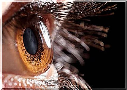
Keratoconus is an ocular condition that specifically affects the cornea, the transparent, dome-shaped membrane that forms the front part of the eye, covering the iris. The function of the cornea is to focus the light rays for good vision.
It is a progressive disease that changes the shape of the cornea, turning its concave shape into that of a cone. In addition, the cornea also becomes thinner and this changes the way the light rays enter. This means that the eye cannot focus properly, leading to blurred vision.
Keratoconus usually affects teenagers and young adults. It has a significant impact on the quality of one’s life as it greatly affects eyesight. In today’s article, we’ll explain everything you need to know about this condition.
What is Keratoconus?

As we mentioned above, the normal shape of the cornea is dome-like. This shape is important because it ensures that the light rays hit the retina just right so that the eye can perform the vision process properly.
Keratoconus gradually modifies the cornea, changing the dome shape and becoming conical. It is a non-inflammatory pathology that can occur in one eye or in both eyes.
These changes in corneal morphology prevent the light rays from being reflected correctly. As a result, it distorts and distorts vision. In addition, a person can develop both nearsightedness and astigmatism.
- Myopia or nearsightedness is a defect that blurs distant objects.
- Astigmatism is when your eye perceives objects as blurry or distorted, both near and far.
What other symptoms can occur with keratoconus?
Many people with this condition also have extreme sensitivity to light. They are also forced to have their glasses adjusted regularly. Symptoms usually vary over time.
Redness and a lot of discomfort when wearing contact lenses are a common symptom. It normally takes a long time for the condition to progress, so the latter symptom is a bit more unlikely.
However, there are cases where keratoconus can become much more serious. The eyes may suddenly deteriorate and the cornea may begin to develop the process of fibrosis or scarring. When this happens, the cornea becomes less transparent and vision deteriorates even more.
What are the causes?
We still don’t know exactly what causes the occurrence of keratoconus. However, scientists believe that many factors are involved in its development.
First, it seems that genetics plays an important role, as many of the people who have it also have a family history. It also has to do with Down and Ehlers-Danlos syndromes.
Also, factors such as minor eye injury, persistent scratching and eye irritation appear to be related, as well as continuous exposure to the sun’s rays.
How do doctors diagnose and treat it?
A keratoconus diagnosis is relatively easy (link in English). Normally, it is sufficient for an ophthalmologist to perform a complete eye examination. In addition, they can perform other tests to observe a particular pathology in more detail.
For example, they may perform a light scan, a corneal topography, or a kerometry test, which can measure the curvature of the cornea. The treatment of keratoconus depends on the entire situation.
Only the ophthalmologist can determine which method is most suitable for each patient. They can treat many mild cases with contact lenses. However, more complex cases may require a corneal transplant.
Summarized
Keratoconus is a progressive condition that deteriorates the cornea and prevents it from focusing light rays properly. This causes the vision to fade. It usually affects young people. When in doubt, the best option is to see an ophthalmologist for a full examination.


